Developing Light-Activated Tissue Adhesive Patch for Rapid, Watertight Neurosurgical Sealing
Durotomy, a tear in the dura mater, is a common complication in neurosurgical procedures. The dura mater is a tough protective membrane that surrounds the brain and spinal cord, safeguarding the central nervous system. When this membrane is damaged, cerebrospinal fluid (CSF) can leak, leading to complications such as delayed wound healing, low-pressure headaches, and serious infections. Preventing CSF leakage, therefore, requires a reliable, watertight closure of the dura mater.
Tissue adhesives are increasingly explored as alternatives to suturing for dural closure because they offer simpler and faster application. However, many existing glue-based sealants suffer from excessive swelling, leading to mass effect, and unwanted tissue adhesion, which can lead to postoperative complications. To address these limitations, researchers have investigated Janus tissue patches, which feature two distinct surfaces—one that adheres strongly to tissue and another that prevents unwanted adhesion. Unfortunately, most existing Janus patches rely on multiple materials and complex, multi-step fabrication processes, limiting their practical use.
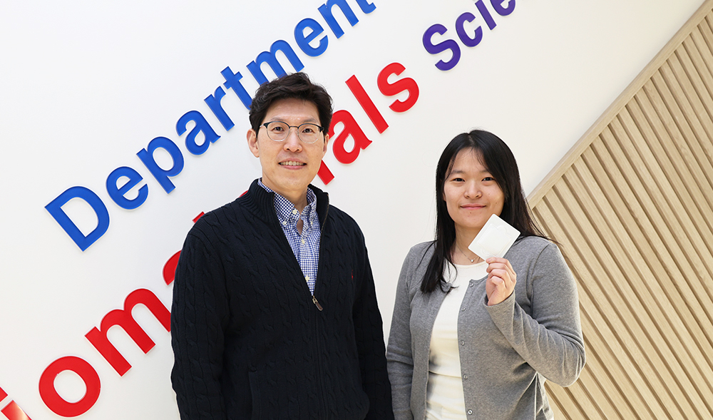
In a breakthrough study, a research team from South Korea led by Professor Seung Yun Yang from the Department of Biomaterials Science at Pusan National University has developed an innovative light-responsive, monolithic Janus dural patch using photocurable hyaluronic acid (HA) through a simple approach. “Made from natural biopolymer hyaluronic acid, our dural patch provides strong wet adhesion, along with a lubricating surface that prevents unwanted tissue adhesion, after exposure to non-toxic visible light,” explains Prof. Yang. Their study was made available online on December 16, 2025, and published in Volume 527 of Chemical Engineering Journal on January 01, 2026.
The researchers selected HA because of its excellent biocompatibility as well as its intrinsic anti-adhesive and lubricating properties. To enable light activation, HA was chemically modified with photocrosslinkable groups—methacrylate (MA) and 4-pentenoate (PA). The resulting HA-based solution was then lyophilized to form a patch with two distinct surfaces: a dense surface with a high polymer concentration and a porous surface with a lower polymer concentration. To further enhance conformal adhesion to wet tissues, the patch was compressed to a thickness of approximately 0.2 mm.
Laboratory tests showed that the patch could fully seal the wounds within five seconds using low-energy visible light. The dense outer surface exhibited strong wet adhesion, achieving high burst pressure and approximately 50% lower friction than conventional dural sealants. Notably, the adhesion strength was up to ten times higher than that of commercially available tissue adhesives. Meanwhile, the porous surface efficiently absorbed fluids and helped prevent unintended tissue adhesion. The patch also demonstrated minimal swelling and a reduced mass effect—less than 200% swelling and an approximately 0.1 g increase in weight—along with high stretchability, flexibility, and excellent biocompatibility.
The team also tested the developed patch in a rabbit durotomy model, where it achieved rapid and effective dural closure without causing damage to the surrounding skull, dura mater, or brain tissue. This photocurable dural patch has been transferred to the biotech company SNvia, where large-scale manufacturing facilities for photocrosslinkable HA have been established. Nonclinical studies are scheduled to be completed in the first half of 2026, and preparations are underway for submission of a medical device clinical trial application to the Ministry of Food and Drug Safety (MFDS, South Korea) within the same year.
“Our technology will allow surgeons to seal wounds rapidly, significantly reducing the risk of postoperative cerebrospinal fluid leakage,” remarks Prof. Yang. “While photocuring technologies are already well established as industrial platform technologies, ensuring safety has remained a major challenge in medical applications. This study is significant in that it provides practical evidence for the clinical applicability of photocrosslinkable hyaluronic acid (HAMA-PA). Its strong adhesion to wet tissues also opens new possibilities for drug-delivery patches, cell-laden constructs, and artificial tissues.”
Overall, this innovative dural patch holds great potential for use in diverse applications requiring rapid, watertight sealing.
- Author (Pusan National University): Seung Yun Yang (Department of Biomaterials Science)
- Title of original paper: A monolithic Janus dural sealant with adhesive and lubricant surfaces
- Journal: Chemical Engineering Journal
- Web link: https://doi.org/10.1016/j.cej.2025.171881
- Contact e-mail: syang@pusan.ac.kr
 953양승윤교수.jpg
(441KB)
953양승윤교수.jpg
(441KB)